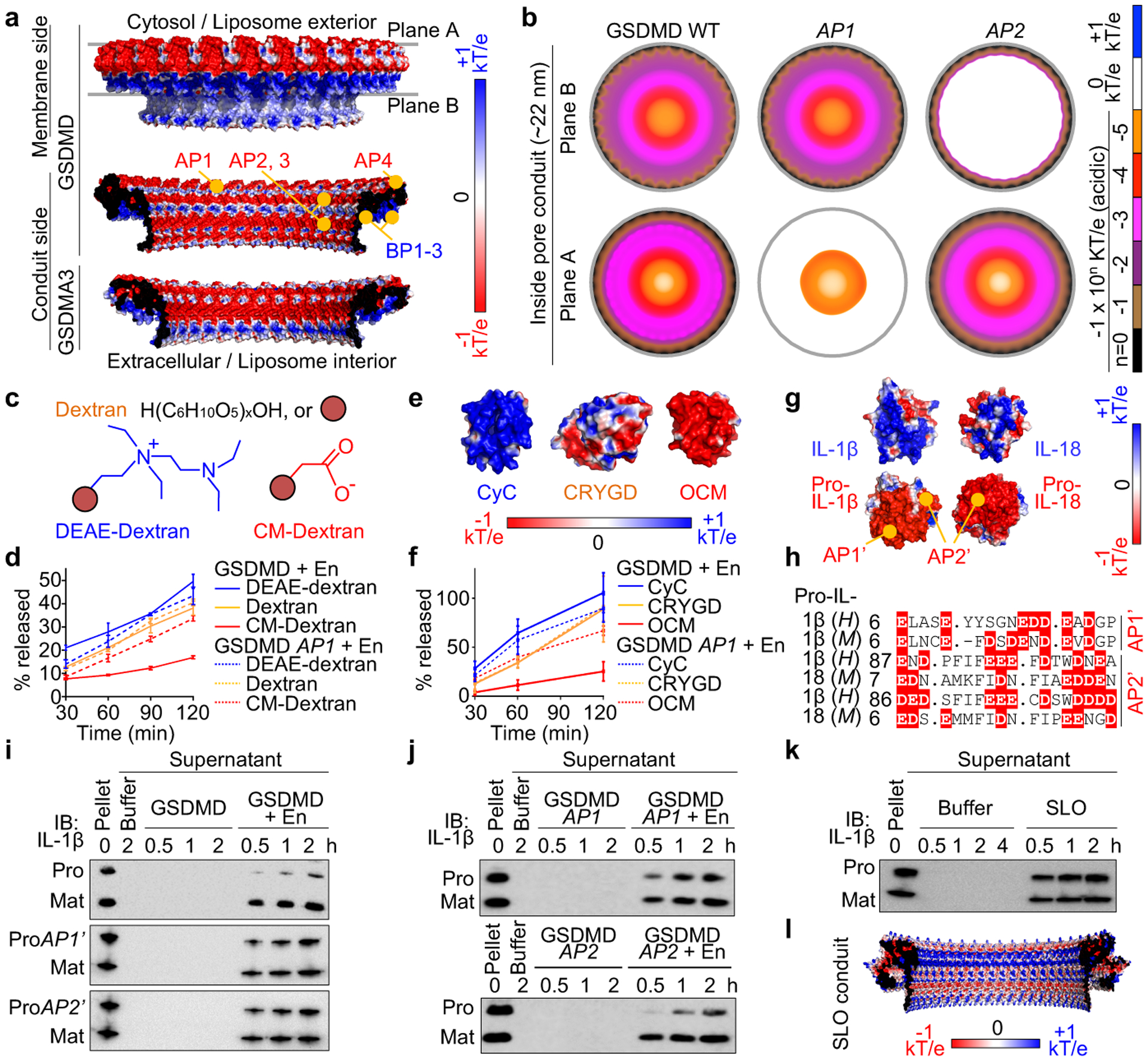
Fig. 3 |. Pore conduit and cargo transport.

a, Electrostatics surface (−1 to +1 kT/e) of the GSDMD pore, with the membrane-bound side formed by BPs and the solvent-exposed side formed by APs. The GSDMA3 pore conduit is similarly acidic. b, Negative electrostatic coverage of the GSDMD pore conduit. Modelled AP1 (5 D/E to A) and AP2 (2 D/E to A) pores have locally neutral conduits. c, d, Cartoons (c) and release of dextran cargoes from liposomes perforated by GSDMD, quantified by FITC fluorescence (n = 3 biological replicates) (d). e, f, Electrostatics surfaces of three protein cargoes (PDB: 2B4Z, 1H4A, 1TTX) (e) and their release from liposomes permeabilized by GSDMD, evaluated by immunoblot quantification (n = 3 biological replicates) (f). g, h, Basification of IL-1β and IL-18 through caspase-1-induced maturation (g) and AP’s of the precursors shown by aligned sequences (h). H: Human. M: Mouse. Dashes: Gaps. Dots: Strings of omitted residues. i, j, Release of pro- (WT and AP’-mutant) and mature and IL-1β from liposomes permeabilized by WT GSDMD (i) and the reciprocal experiments (j). ProAP1’: 8 D/E to K. ProAP2’: 11 D/E to K. k, Release of pro- and mature IL-1β from liposomes perforated by SLO. l, Electrostatics surface of the modelled SLO pore conduit. Data shown in d and f are mean ± s.d.. Data shown in i-k are representative of two independent experiments.
