Figure 1.

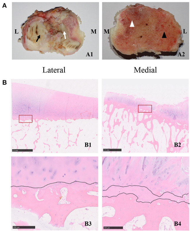
(A,B) Histological characterizations of the cartilage and subchondral bone in the tibial plateau of OA specimens. (A1) The characterizations of the cartilage in different compartments. The black arrow indicates the less-worn cartilage, while the white arrow indicates the severe-worn cartilage in the medial compartment. M indicates the medial compartment of the knee joint, and L indicates the lateral compartment of the knee joint. (A2) The characterizations of the subchondral bone in different compartments. The white arrowhead indicates the severe-degenerative subchondral bone, the black arrowhead indicates the mild-degenerative subchondral bone in the lateral compartment. M indicates the medial compartment of the knee joint, and L indicates the lateral compartment of the knee joint. (B1) Representative hematoxylin and eosin staining images of specimens in the lateral compartment of knee joint. The scale bar represents 2.50 mm. (B2) Representative hematoxylin and eosin staining images of specimens in the medial compartment of the knee joint. The scale bars in B1,B2 represent 2.50 mm. The bottom two panels (B3,B4) are high-magnification images of the red frame in B1 and B2, respectively, the dotted line indicates the tideline. The scale bar represents 250 um. The right panel (B4) is a high-magnification image of the red frame in (B2), and the dotted lines indicate the duplicated tideline. The scale bar represents 250 um.
