Figure 1.
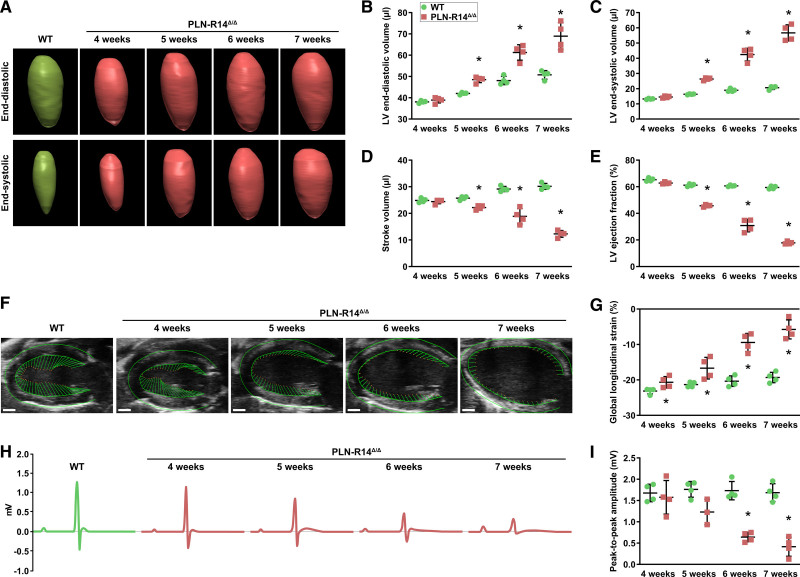
PLN-R14Δ/Δ mice develop progressive dilated cardiomyopathy with heart failure between 4 and 7 wk of age. A through E, Representative 3-dimensional echocardiographic reconstructions of left ventricular (LV) end-diastolic (top) and end-systolic (bottom) volumes of male WT (wild-type; 5 wk old) and PLN-R14Δ/Δ mice (A) with quantification of end-diastolic volume (B), end-systolic volume (C), stroke volume (D), and ejection fraction (E) in WT and PLN-R14Δ/Δ mice at 4, 5, 6, and 7 wk of age (n=4 per group). F and G, Representative long-axis B-mode echocardiographic images of male WT (5 wk old) and PLN-R14Δ/Δ mice with delineation of LV epicardial and endocardial borders showing vectors that indicate direction and magnitude of wall motion (scale bars, 1 mm; F) with quantification of global longitudinal strain in WT and PLN-R14Δ/Δ mice at 4, 5, 6, and 7 wk of age (n=4 per group; G). H and I, Representative average tracings of 1-min ECG recordings of male WT (5 wk old) and PLN-R14Δ/Δ mice (H) with quantification of peak-to-peak amplitudes in WT and PLN-R14Δ/Δ mice at 4, 5, 6, and 7 wk of age (n=4 per group, except n=3 for 5-wk-old PLN-R14Δ/Δ mice; I). Data are presented as mean±SD. *P<0.05 vs age-matched WT mice (Mann-Whitney U test).

