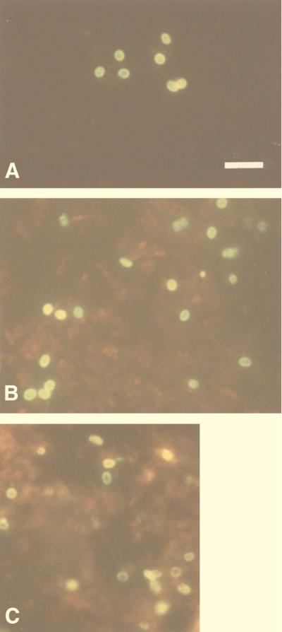
FIG. 1.
Purified whole spores of E. bieneusi stained by indirect immunofluorescence with MAbs 6E52D9 (A) and 3B82H2 (B) in ascitic fluid. MAbs recognize antigens localized in the spores walls. (C) Formalin-fixed smear of a fecal sample, from one of the 14 patients with microsporidia, reacted with a 1:512 dilution of MAb 6E52D9. Note the bright fluorescence of spore walls. Bar = 5 μm.

