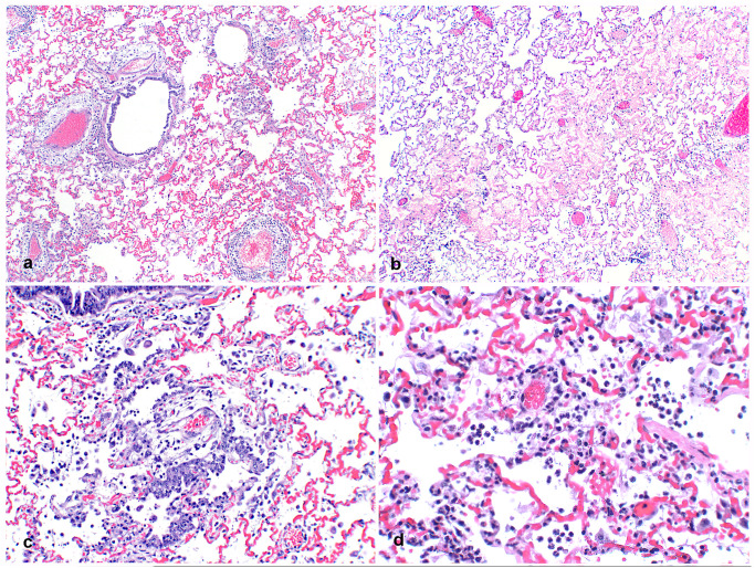
Fig 2. Pulmonary histopathology of SARS-CoV-2 infected mink.
A. Lung from an adult mink with large cuffs of mononuclear inflammatory cells and edema multifocally surrounding pulmonary vessels. 20x H&E. B. Alveolar spaces are multifocally filled with eosinophilic edema fluid. 40x H&E. C. Bronchioles are lined with proliferative, slightly disorganized hyperplastic epithelium and type II pneumocyte hyperplasia is present in alveoli associated with increased intra-alveolar inflammation. 100x H&E. D. Neutrophils, fewer macrophages, and strands of fibrin are multifocally present in alveoli. 200x H&E.

