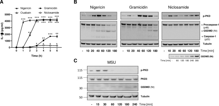
Fig 1. PKD auto-phosphorylation coincides with NLRP3 activation.
(A) IL-1β secretion measured using THP-1 cells primed with 100 nM PMA and stimulated with nigericin (15 μM), ouabain (1 uM), gramicidin (10 uM) or niclosamide (1uM) after the indicated time course. This is one of two experiments with similar results. Statistical significance was calculated with a Student T-test. (B) Stimulation time course using THP-1 cells treated with either nigericin, gramicidin, or niclosamide followed by whole cell lysates analysis by immunoblotting to detect PKD activation (auto-phosphorylation) and NLRP3 inflammasome activation (caspase-1 and gasdermin D cleavage). Tubulin levels were used as loading controls. This is one of two (gramicidin) or three (nigericin, niclosamide) experiments with similar results. An additional experiment with a prolonged time course in the presence of nigericin was implemented to allow for detecting NLRP3 activation (lower panel). (C) Stimulation time course using THP-1 cells treated with MSU crystals at 250 μg/ml followed by whole cell lysates analysis as done in B; this is one of two experiments with similar results. Total PKD3 levels (reported to be the only PKD family member expressed in THP1 cells [33]) were also monitored.

