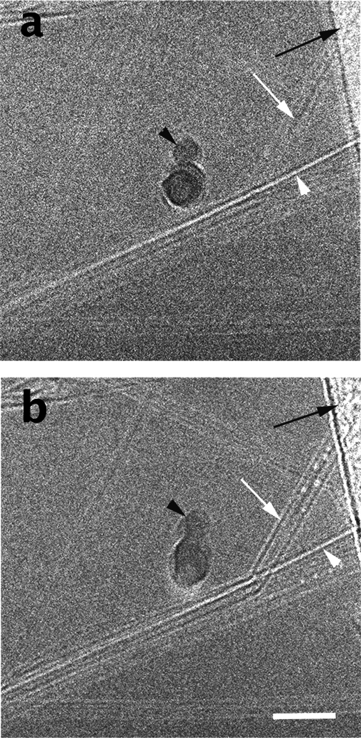
Figure 2.
Vitrified specimen of 0.4 wt % SP10_R BNNTs in CSA. (a) Low-exposure image at about 10 e–/Å2. (b) Same area at ∼40 e–/Å2. Note the changes due to EBRD, including improved image contrast. Black arrows point to the support film, white arrowheads point to an empty BNNT, and black arrowheads point to an ice crystallite. The scale bar corresponds to 50 nm.

