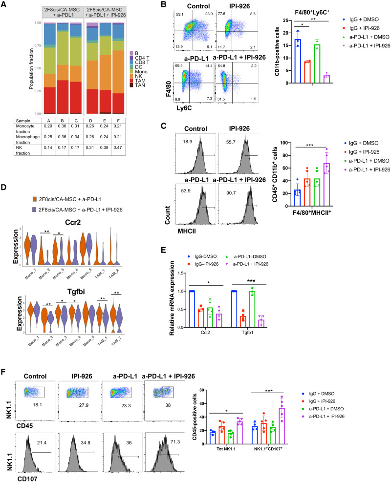
Fig. 6. Dual HHi/a-PD-L1 therapy reduces Ccr2+Tgfbi+ monocytes and macrophages and increases the number of NK cells.
(A) Stacked bar graphs of scRNAseq results showing the proportion of cell types in a-PD-L1–treated or a-PD-L1 + IPI-926–treated 2F8cis/CA-MSC tumor samples. (B and C) Flow cytometry showing the abundance of CD11b+F4/80+Ly6C+ monocytes (B) and the expression of MHCII in CD11b+F4/80+ cells.in the indicated 2F8cis/CA-MSC tumor treatment groups. (D) Violin plots showing the expression of Ccr2 and Tgfbi genes in monocytes and TAM clusters in the indicated tumor treatment groups. (E) RT-qPCR analysis of mRNA expression of Ccl2 and Tgfb1 gene expression in control, a-PD-L1–treated, IPI-926–treated, or a-PD-L1 + IPI-926–treated 2F8cis/CA-MSC tumor samples. (F) Flow cytometric analysis of NK cells, defined as CD45+NK1.1+ cells, and their expression of CD107 in the indicated 2F8cis/CA-MSC tumor treatment groups. Results were analyzed using two-way ANOVA or Wilcoxon signed-rank test. Error bars, SEMs. *P < 0.05, **P < 0.01, and ***P < 0.001.

