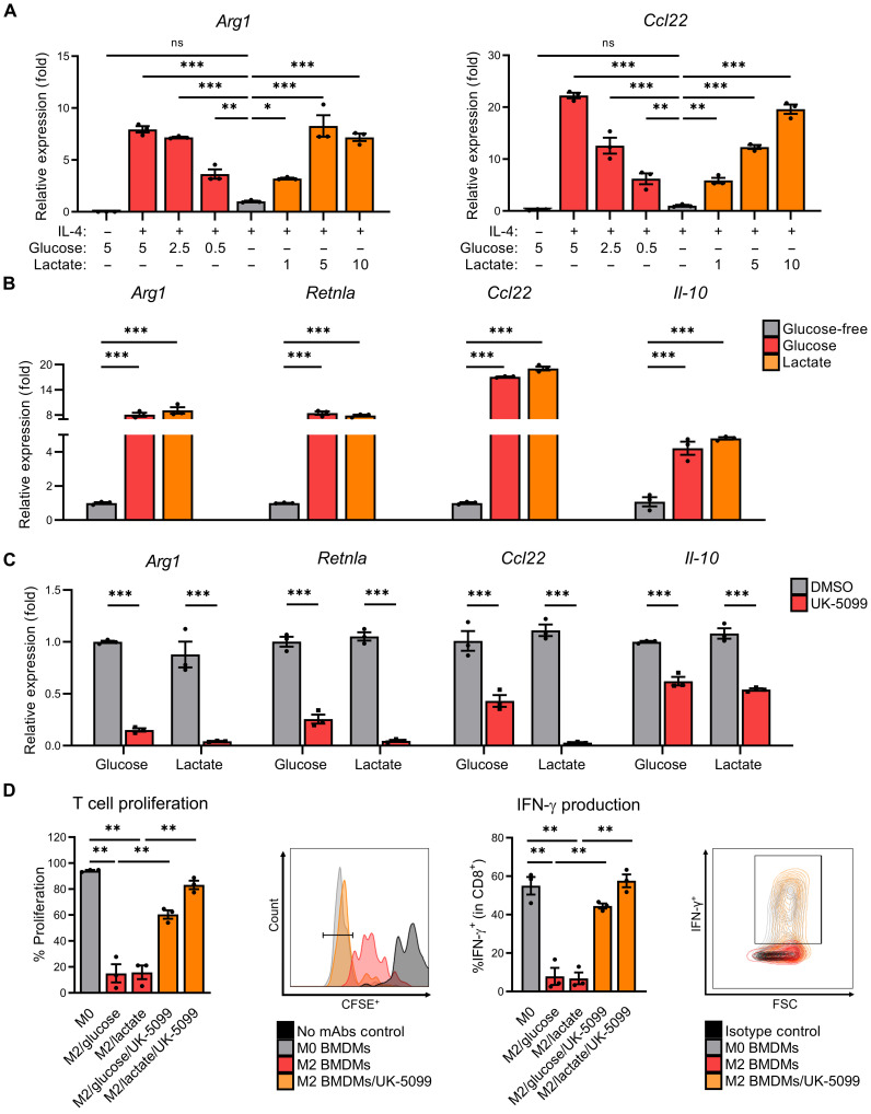
Fig. 1. Macrophages maintain M2 polarization in TME-like conditions through mitochondrial pyruvate/lactate metabolism.
(A) Arg1 and Ccl22 expression of BMDMs starved in glucose-free (GF) media + 10% dialyzed fetal bovine serum (dFBS) (4 hours) before supplementation with glucose or lactate (as indicated), and then polarization with IL-4 (20 ng/ml) for 48 hours. ns, not significant. (B) M2-associated gene expression of BMDMs starved in GF media + 10% dFBS (4 hours) before supplementation with glucose (5 mM) or lactate (10 mM) and polarization with IL-4 (20 ng/ml) for 48 hours. (C) M2-associated gene expression of BMDMs starved in GF media + 10% dFBS (4 hours) before pretreatment ± UK-5099 (25 μM), supplementation with glucose (5 mM) or lactate (10 mM), and polarization with IL-4 (20 ng/ml) for 48 hours. DMSO, dimethyl sulfoxide; mAbs, monoclonal antibodies. (D) Proliferation (left) and interferon-γ (IFN-γ) production (right), with representative images, of CD8+ T cells from splenocytes cocultured for 3 days with BMDMs that were pretreated ± UK-5099 (25 μM) and polarized with IL-4 (20 ng/ml) or vehicle control [phosphate-buffered saline (PBS)] for 24 hours. Data are presented as means ± SEM and represent at least three independent experiments. *P ≤ 0.05, **P ≤ 0.01, ***P ≤ 0.001 by one-way analysis of variance (ANOVA) (A, B, and D) or two-way ANOVA (C) with Tukey’s post-test. CFSE+, carboxyfluorescein diacetate succinimidyl ester–positive.

