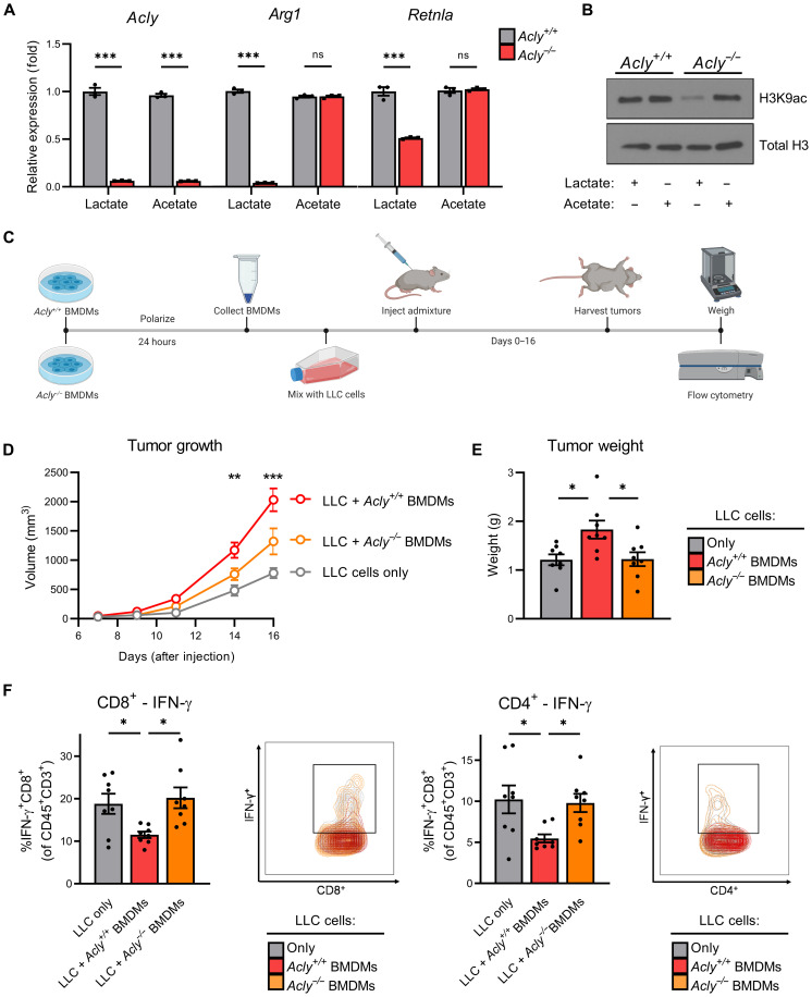
Fig. 4. ACLY is required for maximal M2 macrophage polarization and tumor progression.
(A) M2 gene expression and (B) lysine residue–specific acetylation of Acly+/+ and Acly−/− BMDMs starved in GF media + 10% dFBS (4 hours) before supplementation with lactate (10 mM) or acetate (10 mM) and polarization with IL-4 (20 ng/ml) for 48 (A) or 6 hours (B). (C) Schematic works flow of in vivo tumor admixture model. (D) Growth and (E) weight of tumors consisting of LLC cells only, LLC cells + M2-polarized Acly+/+ BMDMs, or LLC cells + M2-polarized Acly−/− BMDMs. (F) IFN-γ production in CD8+ cytotoxic T cells (left) and CD4+ helper T cells (right) in the respective tumors. Data are presented as means ± SEM of three (A and B) or eight (D to F) replicates. *P ≤ 0.05, **P ≤ 0.01, ***P ≤ 0.001 by two-way ANOVA (B) or one-way ANOVA (D to F) with Tukey’s post-test.

