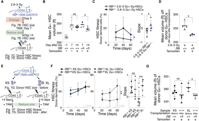
Fig. 7. Reducing cellular size restores HSC fitness.
(A) RB+/+;R26-cre or RBfl/+;R26-cre mice were DNA-damaged (2.8 to 3 Gy) to measure HSC volume (B) and treated with tamoxifen reducing HSC size. Afterward, HSC volume was measured (B) and 700 G0/1 donor HSCs were transplanted into lethally irradiated recipients to measure their fitness (C) and volume (D) in recipients. (B) Mean volume (fl) of G0/1 HSCs from RB+/+;R26-cre or RBfl/+;R26-cre mice after 2.8 to 3 Gy (n = 4) and tamoxifen treatment (n = 5 to 11) as described in (A). Day −1 = before irradiation. Same control and 2.8- to 3-Gy sample as in Fig. 1A. (C) Reconstitution assay: White blood cells (%) derived from donor RB+/+;R26-cre (n; donors = 6, recipients = 11) or RBfl/+;R26-cre (n; donors = 5, recipients = 5) 2.8- to 3-Gy G0/1 HSCs that were treated before transplantation with tamoxifen as described in (A). Slope of reconstitution kinetics was determined. (D) Mean volume (fl) of recipient-derived donor RB+/+;R26-cre (n = 4) or RBfl/+;R26-cre (n = 3) 2.8- to 3-Gy G0/1 HSCs that were treated before transplantation with tamoxifen as described in (A). (E) Experiment schematic: A total of 600 XS- or XL-sized G0/1 HSCs from RB+/+;R26-cre or RBfl/+;R26-cre mice were transplanted into lethally irradiated recipients. Afterward, recipients were treated with tamoxifen to reduce HSC size and to measure fitness (F) and volume (G) of donor G0/1 HSCs in recipients. (F) Reconstitution assay: Percentage (%) of donor-derived white blood cells in recipients and slope of reconstitution as described in (E) (n; donors = 4, recipients XL-RB fl/+ = 6, XS-RB fl/+ = 5, XL-WT = 9, XS-WT = 5). Red arrows indicate tamoxifen treatment. (G) Mean volume (fl) of donor G0/1 HSCs after treatment with tamoxifen from recipients that were reconstituted with XS- or XL-sized G0/1 donor HSCs from RB+/+;R26-cre or RBfl/+;R26-cre mice (n ≥ 3).

