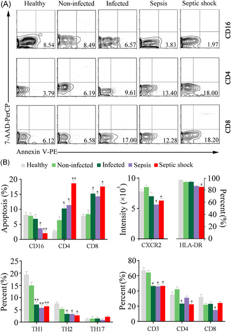
Figure 4.

Variations of the peripheral immune cells in septic patients. (A) Representative histograms illustrating the cell apoptosis of the neutrophils and T lymphocytes. (B) Statistical analysis of flow cytometry results showing the apoptosis and subset changes of immune cells. *p < .05; **p < .01; versus healthy subjects
