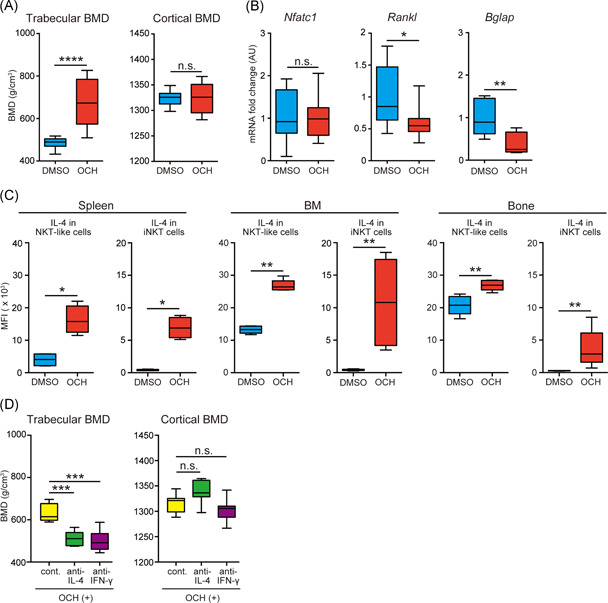
Figure 4.

Administration of OCH suppresses alcohol‐induced osteoporosis in an IL‐4‐dependent manner. Alcohol‐treated B6 mice received intraperitoneal injections of OCH or vehicle every 48 h from 9 to 13 weeks of age. Mice were killed 24 h after the last intraperitoneal injection of OCH or vehicle. (A) Trabecular and cortical bone mineral density (BMD) measured in alcohol‐treated B6 mice that received OCH (red) or vehicle (blue) (8–10 mice/group). The data were pooled from eight independent experiments. (B) Messenger RNA expression of Nfatc1, Rankl, and Bglap in bone from alcohol‐treated B6 mice that received OCH (red) or vehicle (blue) (8–10 mice/group). The data were pooled from eight independent experiments. (C) Intracellular IL‐4 production by natural killer T (NKT)‐like cells and invariant natural killer T (iNKT) cells obtained from the spleen, bone marrow (BM), and bone of alcohol‐treated B6 mice that received OCH (red) or vehicle (blue) (5 mice/group). The data were pooled from four independent experiments. (D) Trabecular and cortical BMD in alcohol‐treated B6 mice that received OCH along with anti‐IL‐4 (green) and anti‐IFN‐γ (purple) neutralizing antibodies. Yellow, isotype matched IgG administration as the control. These antibodies were administered to alcohol‐treated B6 mice at the same time as OCH every 48 h (8–10 mice/group). The data were pooled from four independent experiments. Box plots: horizontal lines of boxes represent the medians, the boxes represent the interquartile ranges, and the whiskers extend to extreme values. *p < .05, **p < .01, ***p < .005, ****p < .001; Mann–Whitney U test. n.s., not significant
