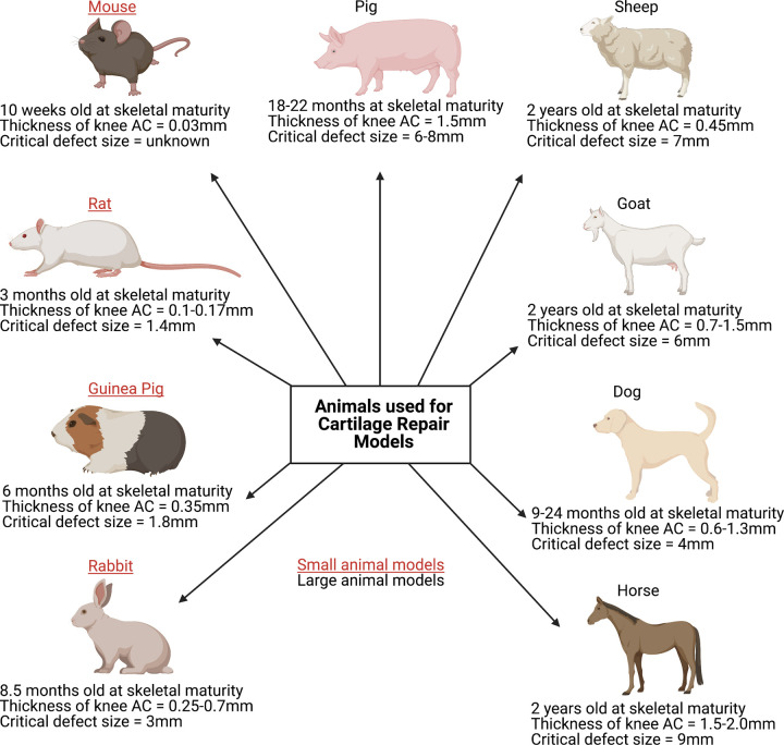
Figure 4. Selected characteristics of animals used for cartilage repair models.
The most commonly used small animal models include the mouse, rat, guinea pig and rabbit, whilst typically the dog, sheep, goat, pig or horse are considered ‘large animal’ models. With numerous models currently available, choosing the most appropriate remains a challenge, although it is vital to note that a single model cannot encompass all of the extensive aspects involved in human cartilage repair [87]. Both small and large animals have their advantages and disadvantages for example; small animal models reach skeletal maturity faster, thereby reducing husbandry costs, experimental durations, drug and housing requirements. However, larger animals present with greater anatomical similarity in regards to the thickness of articular cartilage, joint size and biomechanics to humans. For example, in mice, the average articular cartilage thickness (mm) in the knee joint is ∼0.03 mm, whereas in horses it is ∼1.5–2.00 mm and in humans, it is ∼2.2–2.5 mm [87–91]. Both chondral and osteochondral defects of varying sizes are used in cartilage repair models; all known critical-size defects in the knee joint from the different animal species are displayed here [87,89,92]. (Created using Biorender.com.).

