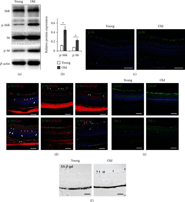
Figure 1.

Increased mTORC1 activity in 24-month-old C57BL/6 retina corresponded with cell senescence. (a) Representative Western blot analysis of total ribosomal protein S6 (S6), phospho-S6 (p-S6), total S6 kinase (S6K), and phospho-S6 kinase (p-S6K) in retinal extracts of 24- (old) and 3-month-old (young) mice. The experiments were repeated three times with one pair of samples at each time. (b) Relative expression of phosphorylated S6 and S6K proteins in control and 24-month-old C57BL/6 retina. Phosphorylation status of each protein was calculated by dividing the intensity of the band of the phosphorylated protein by the intensity of the band of the corresponding total protein. The number from each experiment was averaged, and the error bars represented SE. ∗p < 0.05 by unpaired Student's t-test between 24- and 3-month-old groups. (c) Representative immunofluorescent staining of p-S6 on 24- (old) and 3-month-old (young) mouse retina. (d) Costaining of p-S6 with markers for various retinal cells indicated the activation of mTORC1 in bipolar cells (PKCα, Goα, PKARIIβ), ganglion cells (Brn3a), and amacrine cells (ChAT) but not in Müller glial cells (CRALBP) (arrowheads) of 24-month-old C57BL/6 mouse retina. Arrows indicated aberrant dendritic tip endings of bipolar cells at ONL. (e) Representative immunofluorescent staining showed the activation of Müller glial (GFAP) and microglial (Iba-1) cells in old-aged retina. (f) Senescence-associated-β-galactosidase (SA-β-gal) staining of retinal ganglion cell layer (GCL) (arrows) of the 24-month-old mouse retina. The staining was absent in the retina of young mice. Scale bar: 100 μm.
