Figure 11.

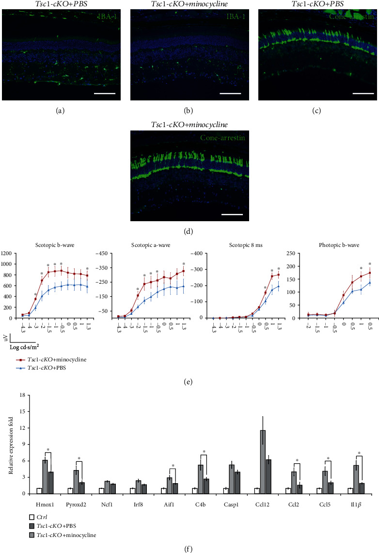
The effect of minocycline on Tsc1-cKO mouse retina. (a, b) Retina sections of Tsc1-cKO retina with or without minocycline treatment were stained with Iba-1 to show the inhibition of microglial cells after minocycline treatment. (c, d) Cone-arrestin staining of the Tsc1-cKO retina with and without minocycline treatment. The minocycline-treated retina showed more arrestin-positive cells, suggesting that the inhibition of microglial cells prevented photoreceptor cell loss. The staining was performed on 6 retina from 6 different animals, 3 in each group. (e) ERG responses of Tsc1-cKO mice with or without minocycline treatment. Three animals in each group were subjected to ERG analysis, and the a-wave, b-wave, and responses at 8 ms after the flash were averaged. Error bars represented SE. ∗ marked significant differences of p < 0.05 by unpaired Students' t-test at the indicated luminance. (f) Real-time PCR analysis of genes associated with oxidative stress (Hmox1, Pyroxd2, Ncf1), microglia activity (Irf8, Aif1, C4b), chemotaxis activity (Ccl12, Ccl2, Ccl5), and inflammatory response (Casp1, Il1β) in Tsc1-cKO retina with or without minocycline treatment. The fold change was relative to untreated- and age-matched control. Six total RNA samples, 3 in each group, were used for PCR analysis. ∗p < 0.05 by unpaired Students' t-test between minocycline- and PBS-treated groups.
