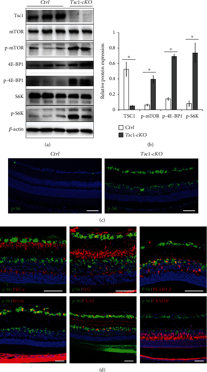
Figure 2.

Characterization of Tsc1-cKO retina. (a) Western blot analysis of Tsc1, mTOR, phospho-mTOR (p-mTOR), 4E-BP1, phospho-4E-BP1 (p-4E-BP1), S6K, and p-S6K in total protein lysates of the Tsc1-cKO and control retina. The experiment was repeated twice. In total, 3 controls and 3 Tsc1-cKO retina samples were analyzed. (b) Relative quantification of protein expression. The density of the bands was normalized against actin for loading control and corresponding total protein for phosphorylated proteins. ∗p < 0.05 by unpaired Students' t-test between control and Tsc1-cKO. (c) Staining of p-S6 indicated the activation of mTORC1 in Tsc1-cKO retina was predominantly at GCL and inner part of the INL layers. Only sporadic staining was found at GCL of control retina. (d) Immunofluorescent staining revealed the localization of p-S6 in bipolar cells (PKCα, Syt2, PKARIIβ), ganglion cells (Brn3a), and some of the amacrine cells (ChAT) of the 3-4-month-old Tsc1-cKO retina. Weak staining was found in Müller glial cells (CRALBP). Scale bar: 100 μm.
