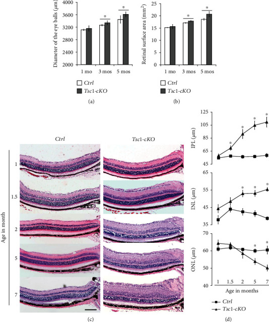
Figure 3.

Histological changes of Tsc1-cKO retina. (a) The diameters of eye balls of 1-, 3-, and 5-month-old Tsc1-cKO and age-matched control mice. (b) Retinal surface area of 1-, 3-, and 5-month-old Tsc1-cKO and control mice. (c) H&E staining of 1-, 1.5-, 2-, 5-, and 7-month-old Tsc1-cKO and control mouse retina showing progressive thickening of the INL and IPL starting at 1.5 months and the thinning of the ONL starting at 3 months of age. (d) The average thickness of IPL, INL, and ONL of Tsc1-cKO and control retina. The thickness was measured on micrographs of H&E staining at about 500 μm away from the center of the optic nerve head. At least 5 eye balls from 5 different animals in each age group of Tsc1-cKO and control were measured, and the results were averaged. Error bars represented SE. ∗p < 0.05 by unpaired Students' t-test between Tsc1-cKO and control group. Scale bar: 200 μm.
