
Figure 4.
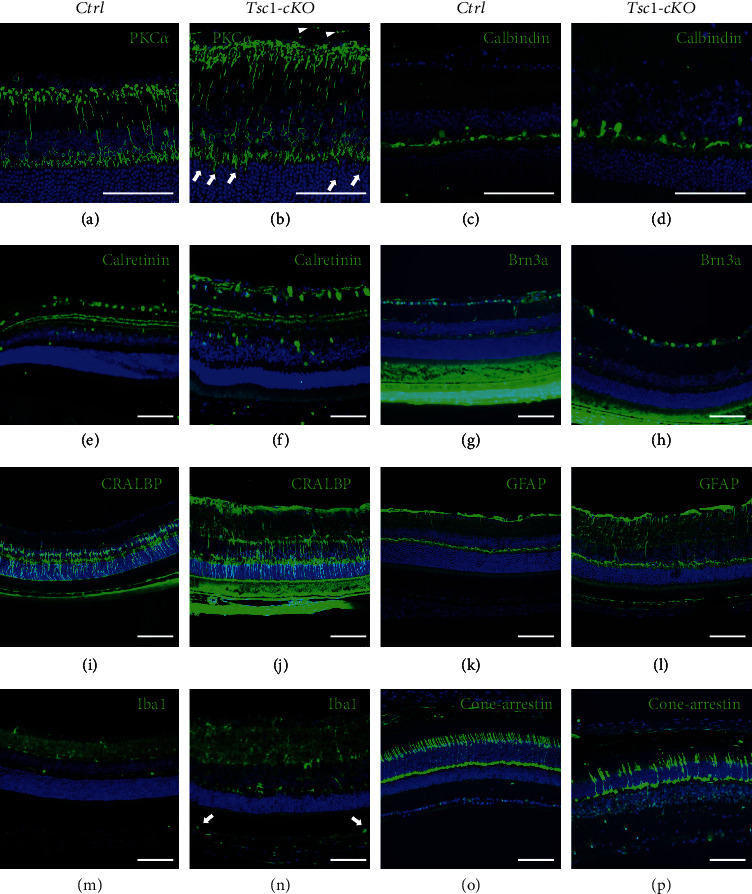
Morphological changes of the Tsc1-cKO retina. (a–l) Immunofluorescent staining of retina from 3-month-old Tsc1-cKO and control mice using antibodies against PKCα for rod bipolar cells (a, b), calbindin for horizontal cells (c, d), calretinin for amacrine cells (e, f), Brn3a for retinal ganglion cells (g, h), CRALBP (i, j), and GFAP (k, l) for Müller glia cells. White arrowheads and arrows in (a) indicated aberrant axon endings and dendritic tips in bipolar cells, respectively. (m–p) Immunofluorescent staining of Iba-1 and cone-arrestin of 7-month-old Tsc1-cKO and control retina to show activated microglia cells (m, n) and dramatically reduced cone photoreceptor cells (o, p). White arrows in (m) indicated Iba1-positive microglia cells in subretinal space. Scale bar: 100 μm.