
Figure 2.
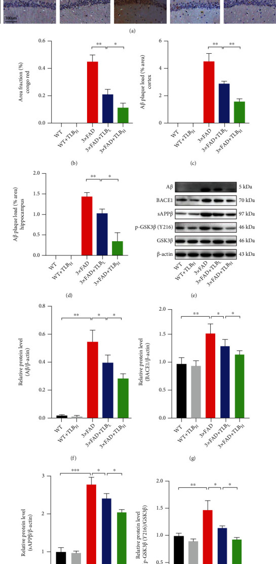
TLB alleviated Aβ deposition in 3×FAD mice. (a) Congo red staining and Aβ IHC staining in the hippocampal area and cortex. (b) Comparison of Congo red staining analysis in the hippocampal area. (c) Comparison of Aβ-positive plaque in the cortex. (d) Aβ plaque load in the hippocampal CA1 area. (e–i) Western blot and quantitative analysis of Aβ, BACE1, sAPPβ, p-GSK3β, and GSK3β in hippocampal tissues. n = 6 per group. ∗p < 0.05; ∗∗p < 0.01; ∗∗∗p < 0.001.