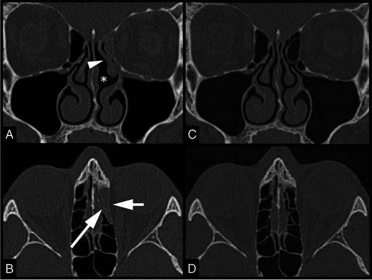
Fig. 1.
CT findings. Coronal (A) CT image shows a lateral deviation of the left middle nasal turbinate (arrowhead), with consequent enlargement of the common nasal meatus (asterisk) and occlusion of the osteomeatal complex. Axial (B) CT image shows opacification and reduced volume of the left anterior ethmoidal cells (long arrow). A mild medial displacement with bowing of the left lamina papyracea is also observed (short arrow). Coronal (C) and axial (D) CT images of 2 years before show that these findings were absent

