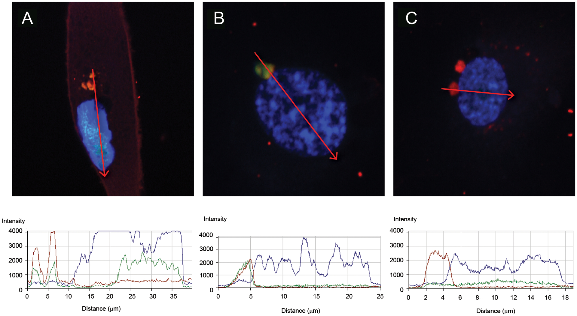
Figure 2: Newly-synthesized RNA from NETs gets internalized by HAECs.

Analysis of the uptake of newly-synthesized RNA from NETs into HAECs by laser confocal microscopy. Shown are representative images of permeabilized HAECs with intracellular newly-synthesized RNA labeled in green, DNA in blue (Hoechst), and neutrophil elastase (A and C) or LL-37 (B) in red. In C, experiment was performed in the presence of RNase A. For each profile image, the fluorescence for each fluorochrome was quantified in the region of the red arrow displayed, and the respective fluorescence quantification is shown in the bottom graphs. The images are representative of at least three different experiments using different SLE and HC samples. Magnification are 126x.
