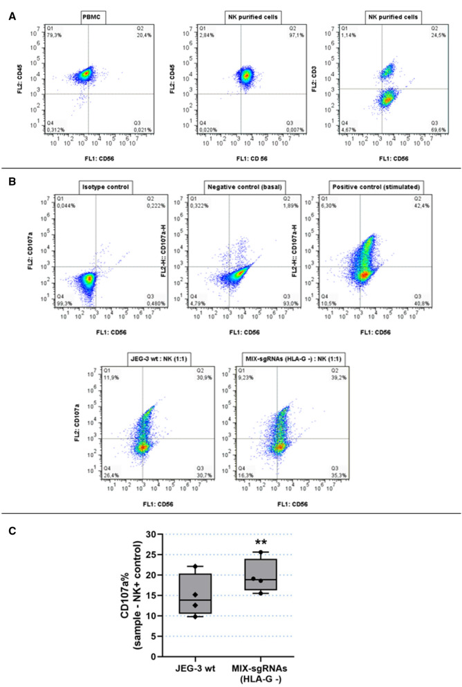
Figure 6.
Degranulation assay, functional analysis of HLA-G wt and HLA-G − JEG-3 cells. (A) Determination of NK cells purification. NK cells were stained with anti-CD45/PE, anti-CD56/BB515 and anti-CD3/PE and compared with PBMC. (B) As a representative assay, NKs were labelled with CD107a/PE and CD56-BB515. The conditions were the following: Isotype control (NK without Ab), negative control (NK basal degranulation), positive control (stimulated NK cells) and NKs co-cultured with wt JEG-3 or with JEG-3/MIX-sgRNAs (HLA-G −). (C) Box plots show the percentage of CD107a + NK cells when co-cultured with wt JEG-3 (HLA-G wt) or with JEG-3/MIX-sgRNAs (HLA-G −). Significant differences are shown with * (p < 0.05) (n = 4 experiments).

