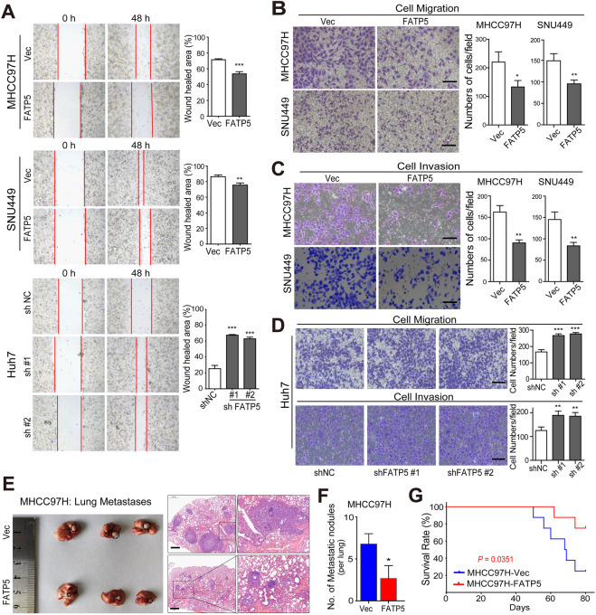
Fig. 2. FATP5 suppresses the invasion and metastasis of HCC cells both in vitro and in vivo.
A Scratch wound-healing assays were performed to evaluate the cell motility in both FATP5-overexpressing and FATP5-silencing HCC cells. After scratching the surface of cell layers, representative images were captured using microscope at 48 h (magnification, 100×) and the wound healed area were calculated according to the remained scratch size at 48 h compared to that area at the beginning. B–D Cell migration and invasion assays were performed to evaluate the migratory and invasive properties. After incubation for 48 h, the migrated and invaded cells were fixed with crystal violet and the cell numbers were microscopically counted. Representative images were shown and results were presented as the average number of cells from 5 independent microscopic fields. Scale bar: 50 μm. All values in (A–D) represented as mean ± SD, *P < 0.05, **P < 0.01, and ***P < 0.001. E, F A nude mouse lung metastasis model was established based on tail vein injection of 1 × 10⁶ MHCC97H-Vec and -FATP5 cells. Representative images (left panel) and H&E stained sections (right panel) showing visible and microscopic metastatic nodules in lung of mice in the MHCC97H-Vec and FATP5-expressing groups (E). The numbers of lung metastatic foci with diameter ≥3 mm in mice were counted and presented as mean ± SD, *P < 0.05 compared with the MHCC97H-Vec group (F). Scale bar: 500 μm. G Comparisons of OS curves in mice injected with either MHCC97H-Vec or MHCC97H-FATP5 cells were analyzed by Kaplan-Meier’s method (P < 0.05 by log-rank test).

