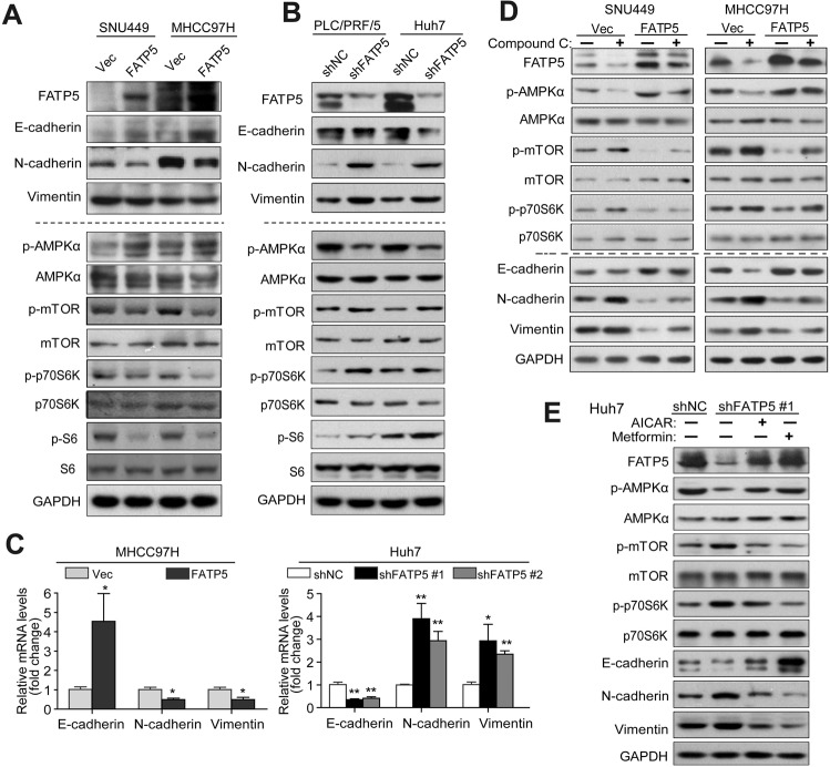
Fig. 3. FATP5 inhibits epithelial-to-mesenchymal (EMT) by regulating AMPK-mTOR-S6K signaling.
A, B Expression of EMT markers (E-cadherin, N-cadherin, and vimentin) and phosphorylation status of AMPKα, mTOR, S6K, and S6 in FATP5-overexpressing cells (SNU449 and MHCC97H transfecting Vec or FATP5) or FATP5 knockdown HCC cells (PLC/PRF/5 and Huh7 cells expressing shNC or shFATP5 #1) were analyzed by western blotting. C The mRNA levels of E-cadherin, N-cadherin, and vimentin in indicated HCC cells transfected with empty vector or FATP5, or infected with LV-shNC or LV-shFATP5 (sequence #1 and #2) were evaluated by qRT-PCR. All mRNA values was normalized to β-actin (ΔCt) and compared with that in their corresponding control cells (ΔΔCt). The fold change was calculated and presented as 2−△△Ct, and the expression levels in MHCC97H-Vec or Huh7-shNC was set as 1. *P < 0.05 and **P < 0.01 (two-tailed t-test). D, E SNU449 and MHCC97H-Vec and FATP5 cells were treated with or without Compound C (10 μM) for 48 h (D). Huh7-shNC and shFATP5 #1 cells were treated with or without AICAR (1.5 mM) or metformin (5 mM) for 48 h (E). Expression of EMT markers and phosphorylation status of AMPK-mTOR-S6K signaling were analyzed by western blotting. For all experiments in (A, B, D, E), GAPDH was used as internal loading control. Abbreviations: p-, phosphorylated.

