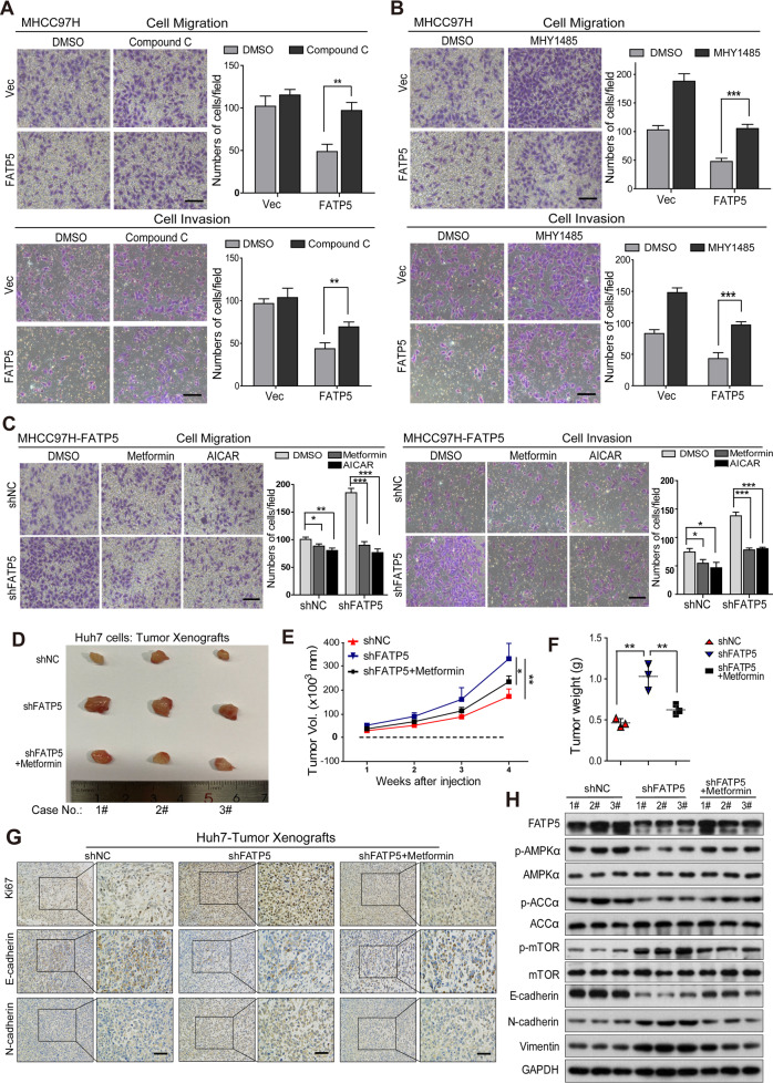
Fig. 5. Activation of AMPK suppresses the EMT process in HCC cells and exhibits antitumor effects in FATP5-deficient xenografts.
A–C MHCC97H cells stably expressing Vec or FATP5 were stimulated with or without Compound C (10 μM) (A) or MHY1485 (10 μM) (B) for 48 h. MHCC97H-FATP5 cells infecting with LV-shNC or LV-shFATP5 #1 were treated with metformin (5 mM) or AICAR (1.5 mM) for 48 h (C). Then the migration and Matrigel invasion assays were performed to measure the migratory and invasive capacities of above indicated HCC cells. The migrated or invaded cells were microscopically counted, and data were presented as the average number of cells from 5 independent microscopic fields. Scale bar: 50 μm. All values in (A–C) expressed as mean ± SD, *P < 0.05, **P < 0.01, and ***P < 0.001. D. Mice injected with 1.5 × 10⁶ Huh7 cells expressing shNC or shFATP5 were treated with control vehicle or daily 4 mg/ml metformin in drinking water for 3 weeks. E, F Xenograft growth conditions from each group (n = 3) were evaluated at indicated time points, and tumor weights were detected after mice were sacrificed. All results were presented as mean ± SD, *P < 0.05 and **P < 0.01. G IHC analyses of Ki67, E-cadherin, N-cadherin in Huh7 shNC or shFATP5-expressing xenografts after treatment of metformin, and representative photographs were shown. Scale bar: 50 μm. H Western blot analyses of the expressions of EMT markers and phosphorylation status of AMPK-mTOR signaling in harvested Huh7-derived tumors as mentioned above. GAPDH was used as internal loading control. #1–3 represented the number of mice in different treated groups.

