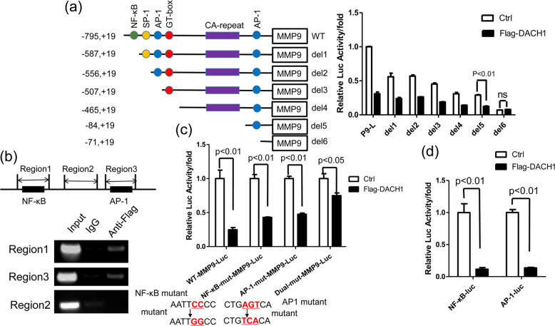
Fig. 3. Determination of DACH1 binding sites in MMP9 promoter.
a HEK 293 T cells were transfected with a series of 5’-deletion constructs of human MMP9 promoter-reporter plasmids together with or without Flag-DACH1. After 24 h, the luciferase activities were measured. The bar graph shows the fold changes in relative luciferase activity normalized against the β-galactosidase activity. There were three independent experiments with three parallel wells each. b ChIP assay showing the binding region of DACH1 in MMP9 promoter. Up, PCR amplified region of MMP9 promoter. Down, HEK 293 T cells were transfected with Flag-DACH1. Cross-linked chromatin was extracted from the cells and subjected to immunoprecipitation with anti-Flag antibody. The DNA regions were amplified by PCR. IgG was used as a negative control. c HEK 293 T cells were transfected with WT-MMP9-luc, NF-κB mut-MMP9-luc, AP-1 mut-MMP9-luc or dual-mut-MMP9-luc together with or without Flag-DACH1. Luciferase activity was measured 24 h after transfection. HEK 293 T cells were transfected with pNF-κB-Luc or pAP-1-luc along with or without Flag-DACH1. Luciferase activity was measured 24 h after transfection. Reporter-gene assay evaluating the regulation of site-specific mutational MMP9 luciferase reporter activity by DACH1 in HEK 293 T cells. The mutation of NF-κB: 5’-AATTCCCC-3’ to 5’-AATTGGCC-3’; the mutation of AP-1: 5’-CTGAGTCA-3’ to 5’-CTGTCACA-3’. d The luciferase activities of pNF-κB-Luc and pAP-1-luc were determined after transfected with Flag-DACH1 or control vector in HEK 293 T cells. There were three independent experiments with three parallel wells each. The data are representative of three independent experiments. Data are presented as means ± SD (P < 0.05, significant; ns, not significant).

