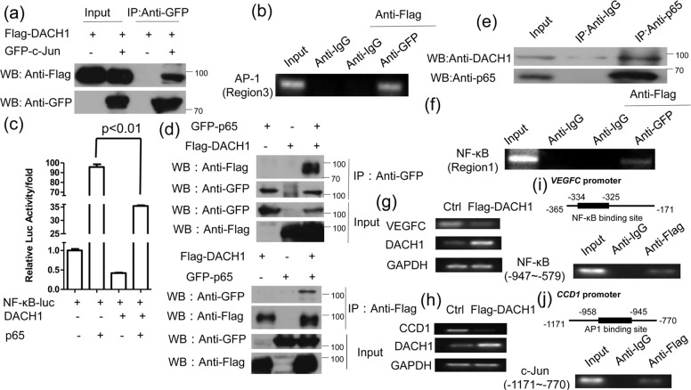
Fig. 4. DACH1 interacts with p65 and c-Jun, respectively.
a HEK 293 T cells transfected with Flag-DACH1 alone or both Flag-DACH1 and GFP-c-Jun were subjected to immunoprecipitation with anti-GFP antibody followed by Western blot with anti-Flag antibody. b HEK 293 T cells were transfected with Flag-DACH1 and GFP-c-Jun. After 24 h of transfection, DNA-protein complex were immunoprecipitated with anti-IgG or anti-GFP antibody, followed by re-immunoprecipitation with anti-Flag antibody. AP-1 binding site (region 3) was amplified by PCR. c HEK 293 T cells were transfected with pNF-κB-Luc and either DACH1 or p65, DACH1 and p65. Luciferase activity was measured 24 h after transfection. d HEK 293 T cells transfected with Flag-DACH1 and GFP-p65 were subjected to immunoprecipitation with anti-GFP antibody followed by Western blot with anti-Flag antibody or vice versa. e ZR-75-30 cells were subjected to immunoprecipitation with anti-p65 or anti-IgG antibody followed by Western blot with anti-DACH1 antibody. f HEK 293 T cells were transfected with Flag-DACH1 and GFP-p65. After 24 h of transfection, DNA-protein complex were immunoprecipitated with anti-IgG or anti-GFP antibody, followed by re-immunoprecipitation with anti-Flag antibody. NF-κB binding site (region 1) was amplified by PCR. There were three independent experiments with three parallel wells each. The data are representative of three independent experiments. Data are presented as means ± SD (P < 0.05, significant; ns, not significant). g, h ZR-75-30 cells were transfected with or without Flag-DACH1, and then subjected to the analysis of CCD1 or VEGFC expression by RT-PCR. i, j Up, the schematic structure of NF-κB binding site in VEGFC promoter or AP1 binding site in CCD1 promoter. Down, ZR-75-30 cells were transfected with Flag-DACH1. Cross-linked chromatin was extracted from the cells and subjected to immunoprecipitation with anti-Flag or IgG antibody. The DNA regions were amplified by PCR. IgG was used as a negative control.

