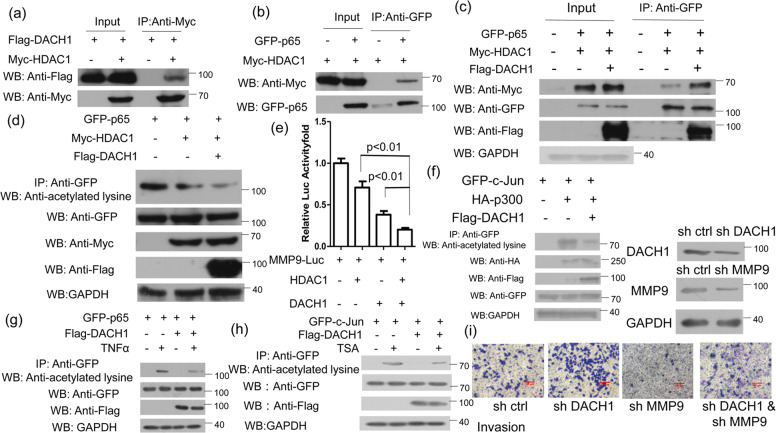
Fig. 5. DACH1 recruits HDAC1 to the NF-kB binding site.
a HEK 293 T cells transfected with Flag-DACH1 alone or Flag-DACH1 and Myc-HDAC1 were subjected to immunoprecipitation with anti-Myc antibody followed by western blot with anti-Flag or anti-Myc antibody. b HEK 293 T cells transfected with GFP-p65 alone or together with Myc-HDAC1 were subjected to immunoprecipitation with anti-GFP antibody followed by Western blot with anti-Myc or anti-GFP antibody. c HEK 293 T cells transfected with GFP-p65 and Myc-HDAC1 along with or without Flag-DACH1 were immunoprecipitated with anti-GFP antibody followed by Western blot with anti-Myc, anti-GFP, anti-Flag antibody. d HEK 293 T cells transfected with the indicated plasmids were subjected to immunoprecipitation with anti-GFP antibody followed by Western blot with anti-acetylated lysine antibody. All cells were treated with TNFα (40 ng/ml) for 4 h before harvest. TNFα was used to activate the NF-κB signaling pathway and promote the acetylation level of p65. e HEK 293 T cells were transfected with MMP9-luc and either HDAC1 or DACH1, HDAC1 and DACH1. f HEK 293 T cells were transfected with the related plasmids for 24 h, and then the cell lysis were subjected to immunoprecipitation with anti-GFP antibody followed by western blot with anti-acetylated lysine antibody. g HEK 293 T cells were transfected with GFP-p65 along with or without Flag-DACH1. Cells were treated with or without TNFα (40 ng/ml) for 4 h before harvest. The lysis were subjected to immunoprecipitated with anti-GFP antibody followed by anti-acetylated lysine antibody. h HEK 293 T cells transfected with the related plasmids for 24 h and then the cells treated by TSA (1 μM) for 6 h before harvest were immunoprecipitated with anti-GFP antibody followed by western blot with anti-acetylated lysine antibody. i MCF7 cell invasion assays was performed in 24-well chambers with Matrigel. Cells were transfected with sh Ctrl, sh DACH1, sh MMP9 or sh DACH1 and sh MMMP9, and then inoculated into the upper chamber. After cultured 24 h, cells on the lower surface of the filter were stained and photographed. The scale bars stand for 100 μm. Luciferase activity was measured 24 h after transfection. Experiments were performed in triplicates and the data are representative of three independent experiments. The data are representative of three independent experiments. Data are presented as means ± SD (P < 0.05, significant).

