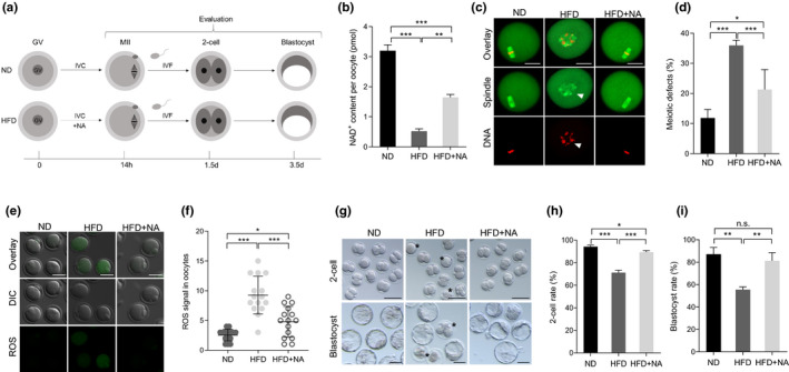
FIGURE 5.

In vitro supplementation of NA improves the developmental potential of oocytes and early embryos from obese mice. (a) The schematic diagram of the experimental procedure. (b) Quantitative analysis of NAD+ content in ND, HFD, and HFD + NA oocytes (n = 150 for each group). (c) ND, HFD, and HFD + NA oocytes were stained with α‐tubulin to visualize spindle (green) and counterstained with propidium iodide to visualize chromosomes (red). Representative confocal sections are shown. Arrowheads indicates the disorganized spindle and misaligned chromosomes. Scale bars: 30 μm. (d) Quantification of ND (n = 128), HFD (n = 120), and HFD + NA (n = 103) oocytes with spindle/chromosome defects. (e) Representative images of CM‐H2DCFDA fluorescence (green) in ND, HFD, and HFD + NA oocytes. Scale bar: 50 μm. (f) Quantification of the levels of ROS in oocytes. Each data point represents an oocyte (n = 15 for each group). (g) Representative images of 2‐cell and blastocyst embryos derived from ND, HFD, and HFD + NA oocytes. Asterisks indicate the abnormal HFD embryos. (h‐i) The percentage of embryos that successfully progressed to the 2‐cell and blastocyst stage during in vitro culture (n = 73 for ND, n = 67 for HFD, and n = 67 for HFD + NA). Data are expressed as the mean ± SD from three independent experiments. Statistical analyses were performed with one‐way ANOVA with Tukey's post hoc test. *p < 0.05, **p < 0.01, ***p < 0.001. HFD, high‐fat diet; NA, nicotinic acid; ND, normal diet; ROS, reactive oxygen species
