Figure 4.
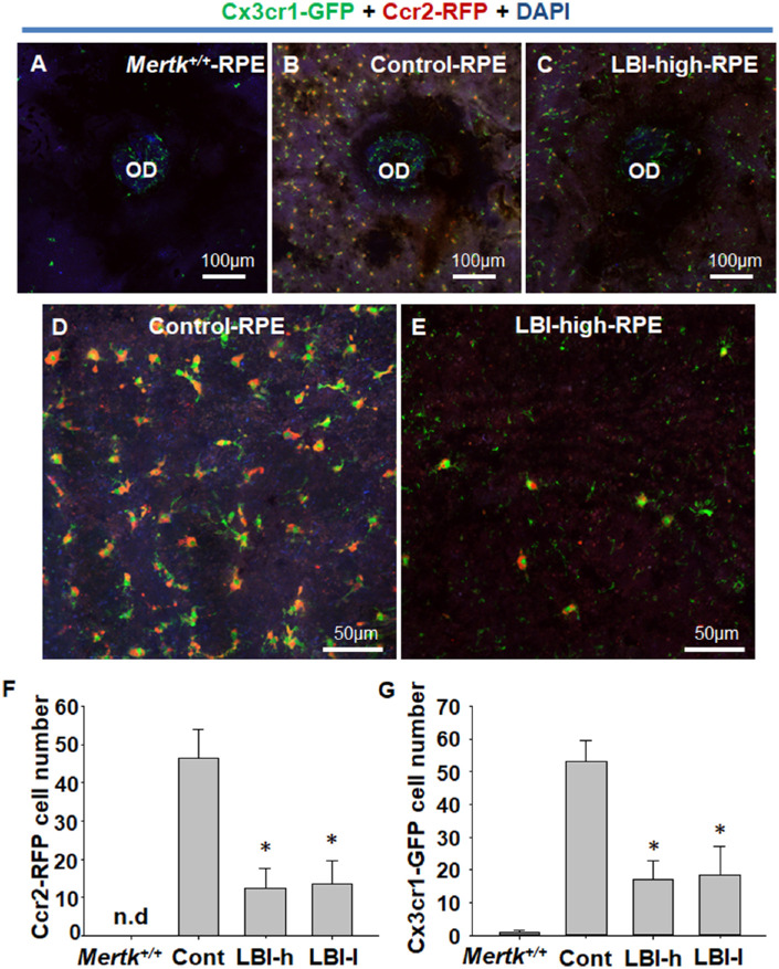
CCR2-RFP– and CX3CR1-GFP–positive cell migration to subretinal space was suppressed by LBI administration. RPE flat mounting was performed to precisely observe the RPE and subretinal space. RPE flat mount of Mertk+/+Cx3cr1GFP/+Ccr2RFP/+ mice (Mertk+/+) is shown as negative control (A). CX3CR1-GFP expression was observed mainly at the OD, and only faint expression was observed at the RPE (A). Control Mertk−/−Cx3cr1GFP/+Ccr2RFP/+ mice showed CCR2-RFP– and CX3CR1-GFP–positive cell migration to the RPE and subretinal space (B and D). LBI-high Mertk−/−Cx3cr1GFP/+Ccr2RFP/+ mice showed suppression of CCR2-RFP- and CX3CR1-GFP-positive cell migration to the RPE and subretinal space (C and E). CCR2-RFP– and CX3CR1-GFP–positive cells of each group (Mertk+/+, n = 4; Control [Cont], n = 12; LBI-high [LBI-h], n = 8; and LBI-low [LBI-l], n = 8) were counted. *P < 0.05 compared with the control group. Separate CCR2-RFP and CX3CR1-GFP images are shown in Supplementary Figure S3.

