Figure 5.
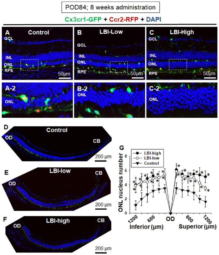
LBI-administered Mertk−/−Cx3cr1GFP/+Ccr2RFP/+ mice showed attenuation of RD at POD 84. LBI-high, LBI-low, and control feed were provided to the mice until POD 84. Severity of RD was assessed using retinal sectioning. Control Mertk−/−Cx3cr1GFP/+Ccr2RFP/+ mice showed severe RD at POD 84 (A and D). Magnified image of the ONL and subretinal space is shown (A-2). Retinal sections of LBI-high (C) and LBI-low (B) Mertk−/−Cx3cr1GFP/+Ccr2RFP/+ mice are shown. Preserved nuclei in ONL were observed in LBI-high (C-2) and LBI-low (B-2) groups compared with those in the control group (A-2). Entire inferior retina (from the OD to ciliary body [CB]) are shown (D, E, and F). ONL nuclei number of each 200 from OD to inferior or superior retina are shown (G). LBI-high (n = 10) and LBI-low (n = 10) groups showed significant attenuation of RD compared with the control group (n = 12). *P < 0.05 compared with the control group.

