Fig. 7.

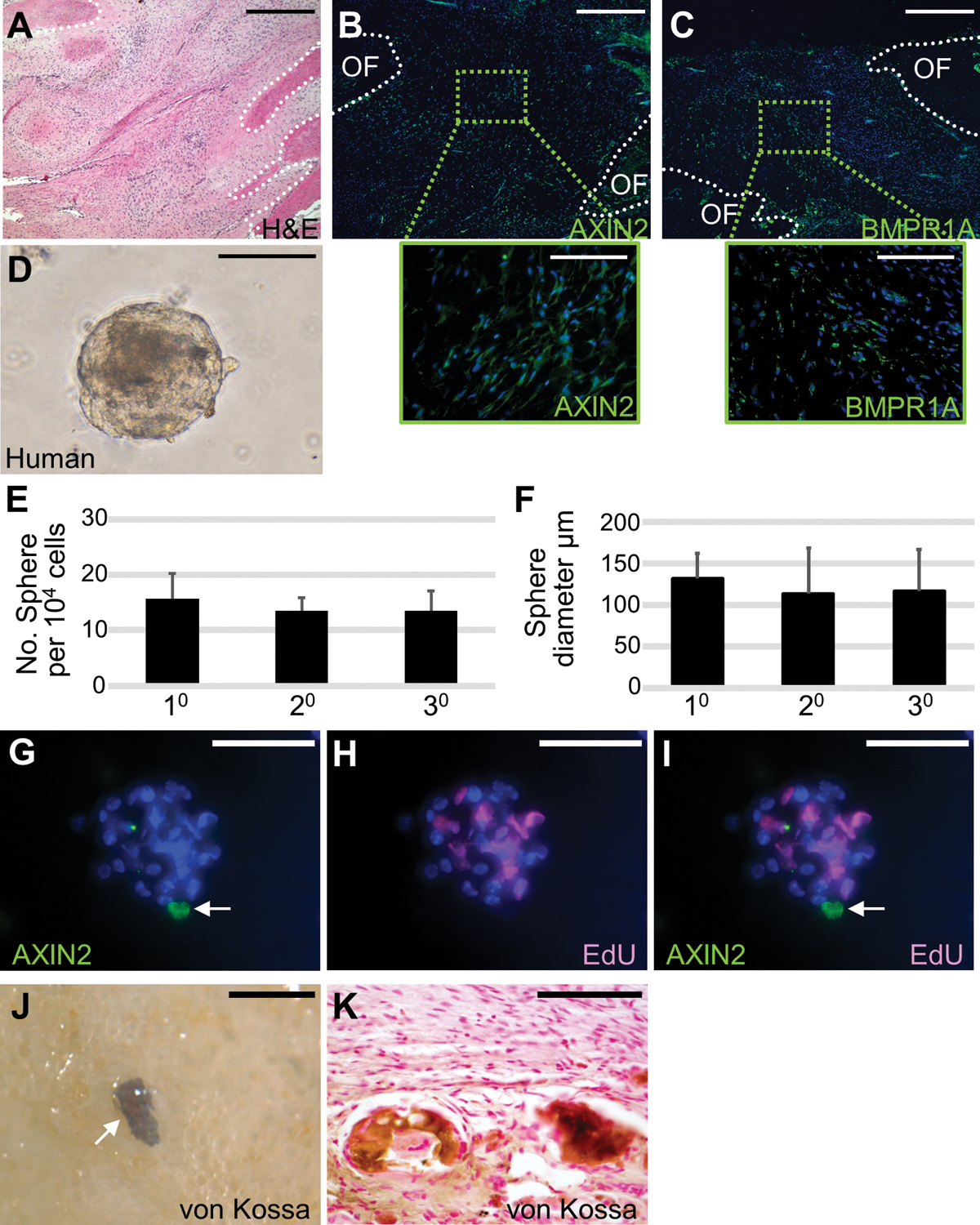
Self-renewal and osteogenic ability of human SuSCs. Sections of the 14-month-old human coronal suture were examined by hematoxylin and eosin staining (A), immunostaining of AXIN2 (B), and BMPR1A (C). Broken lines define the calvarial bones at the osteogenic front (OF). (D) Cells isolated from human suture form spheres in ex vivo culture. Diagrams illustrate the average number (E, n=5, mean ± SD) and size (F, n=5, >15 spheres in each passage, mean ± SD) of spheres formed by the 10, 20, and 30 culture of human suture cells starting with 104 cells for each passage. (G-I) Co-immunostaining of the human sphere after pulse-chase labeling identifies a single AXIN2-expressing cell (arrow) and EdU positive cells undergoing mitotic division. The von Kossa staining in whole-mounts (J) and sections (K) shows ectopic bone formation in the kidney capsule with implantation of human suture cells. Images are the representatives of at least five independent experiments. Scale bars, 500 μm (A-C); 100 μm (B-C insets, D, G-I, K); 300 μm (J).
