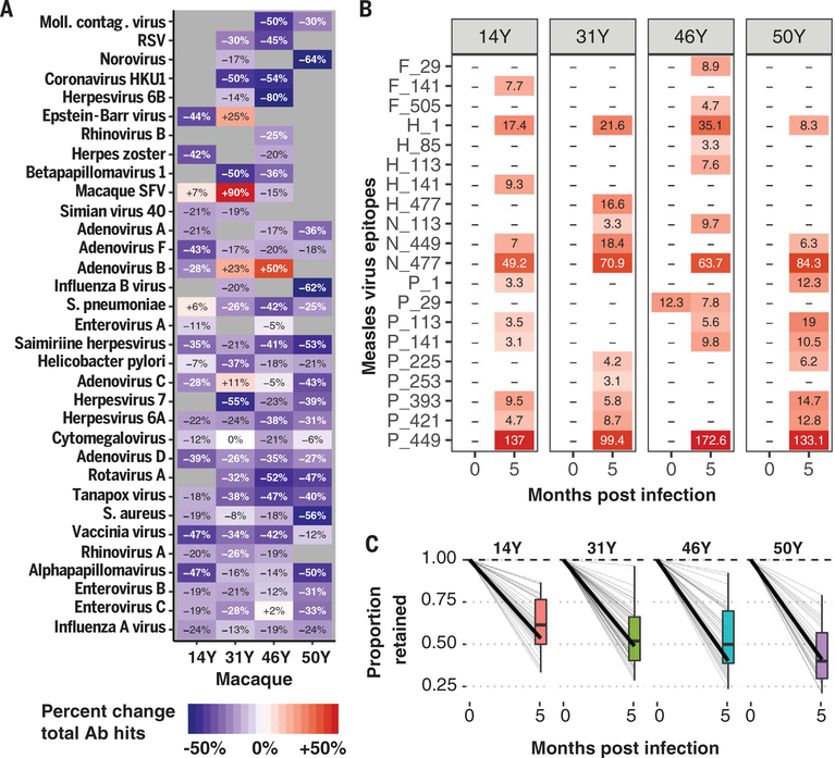
Fig. 5. Measles virus infection in macaques deletes preexisting immune memory.
Four rhesus macaques (14Y, 31Y, 46Y, and 50Y) were infected with the Bilthoven strain of wild-type MV [detailed in (34)], and plasma samples were collected before and 5.1 months after infection. (A) Heatmap showing the percentage change in total epitopes recognized per pathogen in each monkey from time 1 to time 2. Pathogens with >10 unique epitopes recognized at either time point per macaque are shown. The color and text indicate percent change in total epitopes recognized per pathogen species before versus after infection. (B) Heatmap showing the signal strength of anti-measles antibodies (EBS) for each epitope for which at least one monkey developed an antibody. Colors and text of each cell represent the respective EBS values. Letters indicate the MV protein (F, fusion protein; H, hemagglutinin; N, nucleoprotein; and P, phosphoprotein), and the numbers indicate the position along the protein of the first amino acid of the 56-amino-acid peptide. (C) The fractions of epitopes recognized before MV infection that remained 5 months after are shown (0 months after infection is baseline and thus set to 1 for all pathogens). Each gray line indicates a different pathogen, and the dark black line indicates the average across the pathogens. The boxplot summarizes the interquartile range and median fraction retained across the pathogens.

