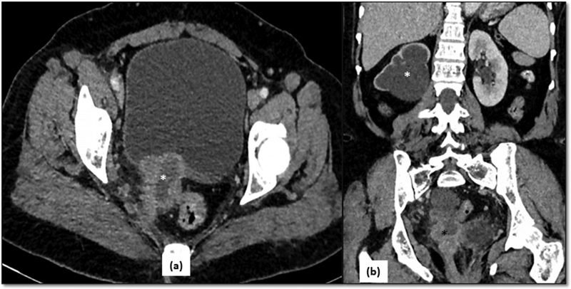
Fig. 4.

CT in stage IVA of carcinoma cervix—( a ) Axial CT image shows an irregular heterogeneous mass in the cervix with central necrosis ( asterisk in a ) infiltrating the urinary bladder anteriorly and reaching up to the lateral pelvic wall posterolaterally on the right side. ( b ) There is upstream right hydronephrosis ( white asterisk ) due to encasement of lower ureter by the mass ( black asterisk ).
