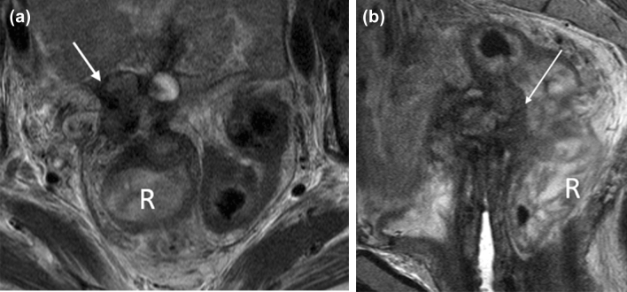
Fig. 12.
62-year-old woman with FIGO stage 4 vaginal cancer. a Axial T2-weighted MR image shows a complex soft tissue mass (arrow) with a cystic component at the superior edge of the vaginal cuff. The mass abuts several small bowel loops, with apparent tethering of surrounding structures. b Sagittal T2-weighted MR image shows that the mass invades the upper rectum (arrow). There is circumferential wall thickening of the upper rectum and sigmoid colon. R = rectum

