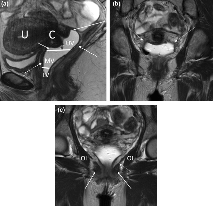
Fig. 8.
Relevant MR anatomy of the vagina. a Sagittal T2-weighted MR image of the vagina (distended with gel) shows the upper (UV), middle (MV), and lower (LV) thirds. The image also shows the anterior and posterior fornices (solid arrows), cervix (C), and uterus (U). The dotted arrows demonstrate the T2-hypointense muscularis layer of the vagina. b Coronal T2-weighted MR image of the vagina shows the lateral fornices of the vagina (solid arrows). c Coronal T2-weighted MR image through the vagina demonstrates the puborectalis (solid arrow), levator ani (dotted arrow), and obturator internus (OI)

