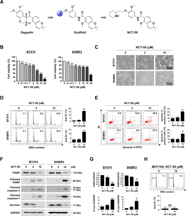
Fig. 1. NCT-58 reduces cell viability and induces apoptosis in HER2-positive breast cancer cells.
A Chemical structure of deguelin and NCT-58. B BT474 and SKBR3 cells were treated with various concentrations of NCT-58 (0.1–20 μM) or DMSO (solvent control) for 72 h. Cell viability was determined by MTS assay (***p < 0.001, n = 4). C Morphological changes of BT474 and SKBR3 cells after treatment with NCT-58 (2–10 μM, for 72 h) as seen through phase-contrast microscopy. D Cells were treated with NCT-58 (2–10 μM) for 72 h and the sub-G1 population was assessed by flow cytometry (***p < 0.01, n = 3). E Early and late apoptotic cells in the presence or absence of NCT-58 were quantified by annexin V/PI staining (right panel, **p < 0.01; ***p < 0.001, n = 3). F Effect of NCT-58 on expression of cleaved-caspase-3, cleaved-caspase-7, cleaved-PARP and survivin in BT474 and SKBR3 cells. G Quantitative graphs of these protein levels (*p < 0.05; **p < 0.01; ***p < 0.001, n = 3). GAPDH was used as an internal loading control. H The sub-G1 fraction of the normal human mammary epithelial MCF10A cells was analyzed through flow cytometry after exposure to 10 μM of NCT-58 for 72 h (NS, not significant, versus DMSO control, n = 3). The results are presented as mean ± SD of at least three independent experiments and analyzed by one-way ANOVA or Student’s t-test followed by Bonferroni’s post hoc test.

