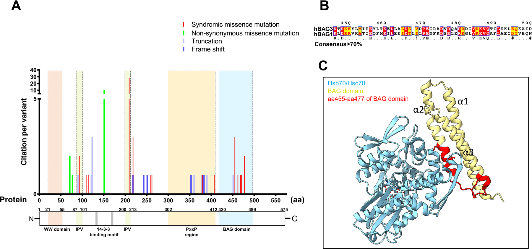
Figure 4. The majority of syndromic mutations in BAG3 are located in the BAG domain.

(A) A diagram that maps the reported variants of BAG3 on the protein sequence. BAG3 variants are color coded and indicate syndromic missense mutations, non-syndromic missense mutations, truncations and frame shifts. (B) Structure-based sequence alignment of BAG domain from BAG3 and BAG1. The sequence ticker of the protein is based on the original sequence of BAG3. (C) The crystal structure of the BAG domain of human BAG1 (yellow) associating with the ATPase domain of bovine Hsp70 (blue, PDB: 1HX1).
