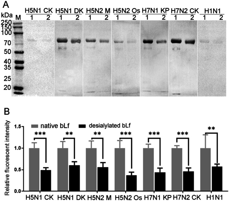
Fig. 2.
Assessing the binding activity of native or desialylated bLf with IAV. A Viral proteins blotting. From left to right: Cy5-labeled viral proteins from H5N1 CK, H5N1 DK, H5N2 Os, H5N2 M, H7N1 KP, H7N2 CK, and H1N1 vaccine in turns. B The relative fluorescent intensity of native or desialylated bLf that bind with IAV. The fluorescent intensity in native bLf was deemed as 1 and compared to that of desialylated bLf. Data was measured from three replications by ImageJ software. M: protein molecular weight markers, lane 1: native bLf, lane 2: desialylated bLf. *P < 0.05, **P < 0.01, ***P < 0.001

