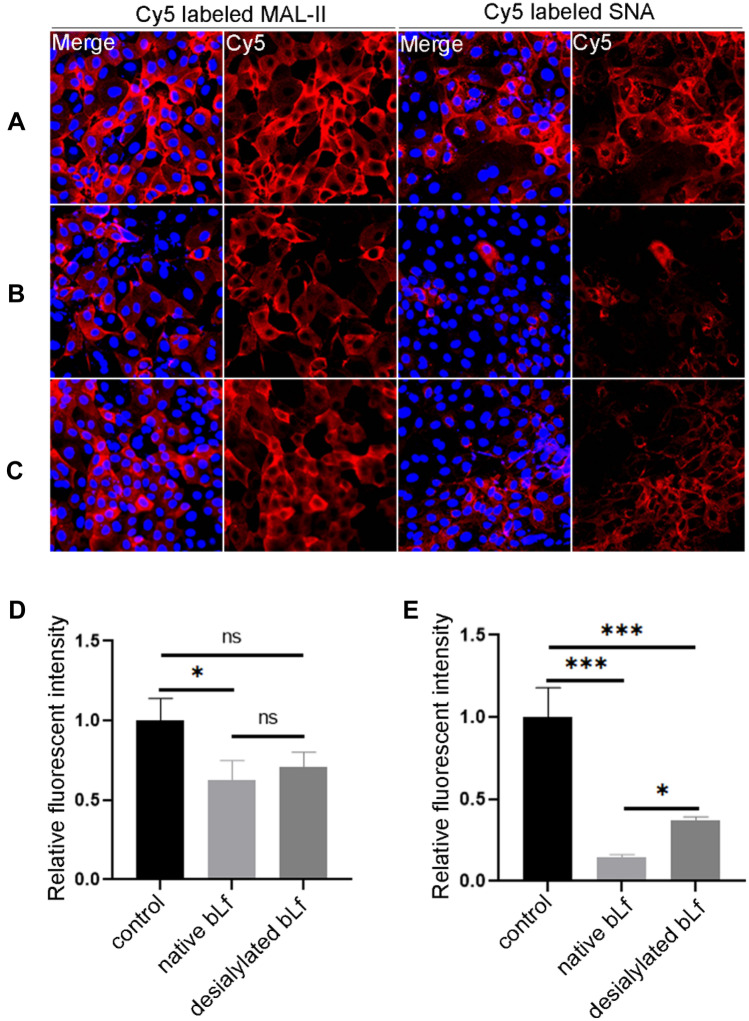
Fig. 3.
Evaluation of the roles of sialylated glycans on bLf against IAV. Histopathologic examination of Cy5 labeled MAL-II and SNA staining performed on MDCK cells. A Control: 25 μg mL−1 of Cy5 labeled MAL-II and SNA were incubated with immobilized MDCK cells. B 20 μg mL−1 native bLf, C 20 μg mL−1 desialylated bLf were added into the incubation solution. The images were acquired under the same conditions for the DAPI merge channel and the Cy5 channel. The relative fluorescent intensity of Cy5 labeled MAL-II D and SNA E bound to MDCK cells were analyzed by ImageJ software. The fluorescent intensity in control was deemed as 1 and compared to that of native or desialylated bLf. Data was measured from three replications. *P < 0.05, **P < 0.01, ***P < 0.001

