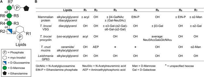
FIGURE 1.
General structure of the GPI anchor and their side chain modifications. (A) Structure of the conserved glycan core with the different side chain modifications. The respective positions of the modifications are indicated by Rx. (B) Comparison of different side chain modifications (Rx) and lipid moieties for selected GPI anchored proteins in mammals, Trypanosoma brucei, Trypanosoma cruzi, and Leishmania. The positions of R1-7 and the lipids in the GPI anchor are indicated in panel (A). The nature and position of linkage of an additional hexose (*) at the first mannose of the GPI anchor of T. cruzi mucins is not yet known (Serrano et al., 1995). This figure was modified from Figure 1 of a review by Fujita and Kinoshita (2010).

