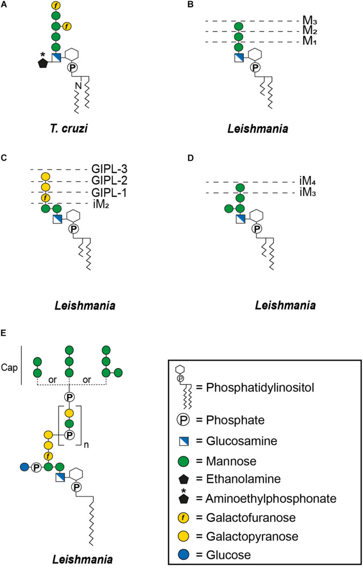
FIGURE 2.
General structures of free GPIs (GIPLs) and LPGs. The dashed lines indicate smaller GIPL species in Leishmania (McConville and Ferguson, 1993). MX indicates the number of mannoses and iM the unusual α1-3 binding of these mannoses. The number of phosphosaccharide repeats (n) of Leishmania LPGs is stage and species specific (Forestier et al., 2014). (A) Trypanosoma cruzi Type-1 GIPL, (B) Leishmania Type-1 GIPLs, (C) Leishmania Type-2 GIPLs, (D) Leishmania hybrid GIPLs, and (E) Leishmania LPGs.

