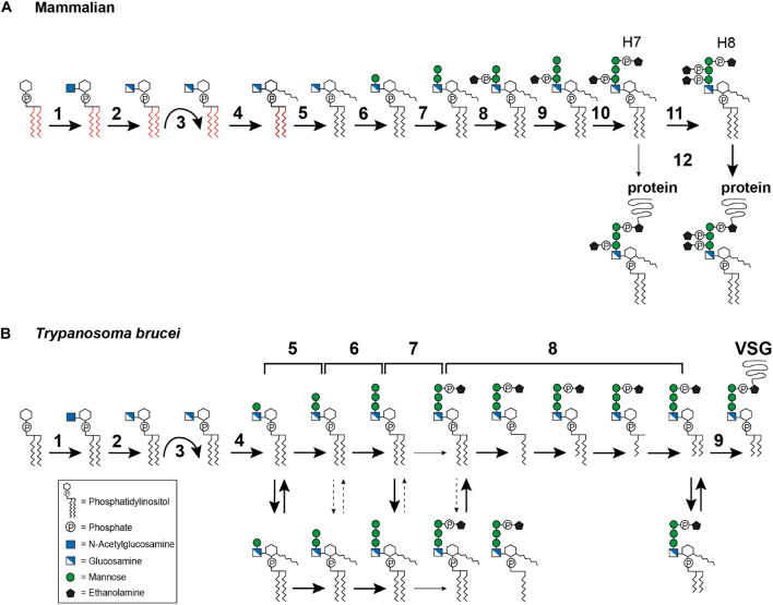
FIGURE 3.
Glycosylphosphatidylinositol biosynthesis pathway up to the point of protein attachment. (A) Mammalian GPI biosynthesis steps at and within the endoplasmic reticulum (ER). The reaction steps are numbered and are described in detail in the text: (1) transfer of N-acetylglucosamine (GlcNAc) to phosphatidylinositol (PI), (2) deacylation of GlcNAc-PI, (3) flipping of GlcN-PI into the ER lumen, (4) inositol acylation, (5) lipid remodeling, visualized by the color change from red to black, (6–7) addition of mannose, (8) addition of ethanolamine phosphate (EtN-P), (9) addition of mannose, (10) addition of EtN-P, (11) addition of EtN-P, (12) attachment of the GPI anchor to the protein. The occasionally observed addition of a fourth mannose is not depicted. (B) GPI biosynthesis steps at and within the ER in Trypanosoma brucei. The reaction steps are numbered and are described in detail in the text: (1) transfer of N-acetylglucosamine (GlcNAc) to phosphatidylinositol (PI), (2) deacylation of GlcNAc-PI, (3) flipping of GlcN-PI into the ER lumen, (4–6) addition of mannose, (7) addition of EtN-P, (8) lipid remodeling, (9) attachment of the GPI anchor to the protein. The broad solid arrows indicate reactions for which direct evidence exists. The dashed arrows indicate conversions that may exist. The light solid arrows indicate reactions that are not frequently observed. The curved arrows indicate the flipping reaction into the ER lumen.

