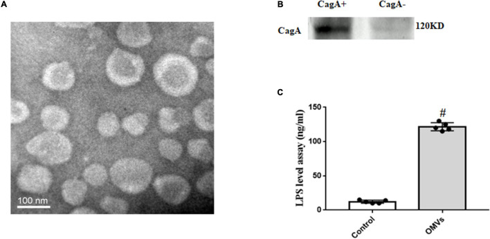
FIGURE 1.
Identification and characterization of OMVs from H. pylori. (A) Electron micrographs of OMVs from H. pylori. Isolated OMVs from H. pylori were fixed with negative staining with Na-phosphotungstate, and observed by transmission electron microscope. (B) CagA expression in H. pylori-derived OMVs (CagA+: OMVs from CagA-positive H. pylori; CagA–: OMVs from CagA-negative H. pylori). (C) LPS levels in H. pylori-derived OMVs and vehicle were determined by ELISA (n = 5, #P < 0.01 vs. control).

