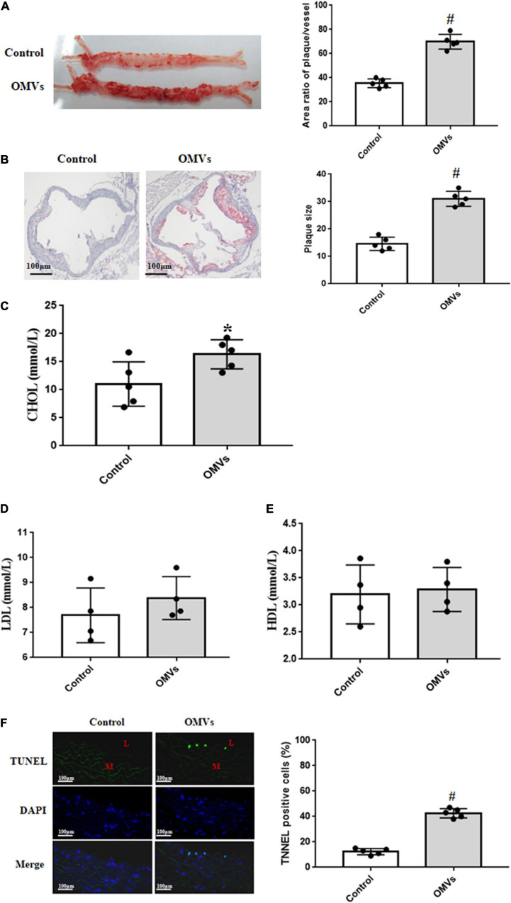
FIGURE 2.
H. pylori-derived OMVs accelerate atherosclerosis plaque formation in ApoE–/– mice. (A) Continuous atherosclerosis plaques were measured by Oil Red O staining in ApoE–/– mice treated with H. pylori-derived OMVs (n = 5, #P < 0.01 vs. control). (B) Cross-section histological analysis were determined by HE staining in ApoE–/– mice treated with H. pylori-derived OMVs (n = 5, #P < 0.01 vs. control). (C–E) The levels of serum lipids, including cholesterol (C, CHOL), low density lipoprotein cholesterol (D, LDL), high density lipoprotein cholesterol (E, HDL), were determined by ELISA in ApoE–/– mice treated with H. pylori-derived OMVs (n = 4 or 5, *P < 0.05 vs. control). (F) Apoptosis were measured by TUNEL staining in arteries from ApoE–/– and control mice. Magnification is × 20; blue, cell nuclei (DAPI staining); green, TUNEL staining; L, lumen; M, media (n = 5, #P < 0.01 vs. control).

