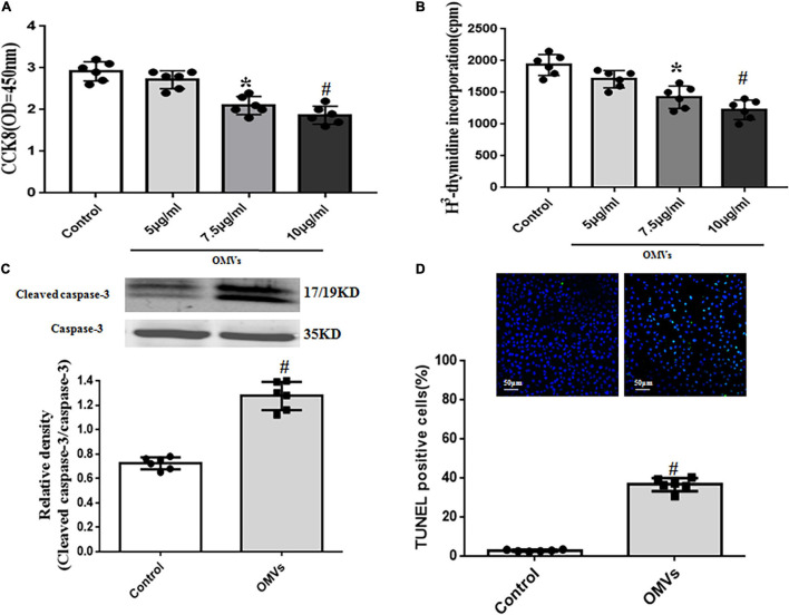
FIGURE 3.
Role of H. pylori-derived OMVs on proliferation and apoptosis of HUVECs. (A,B) Proliferation of HUVECs treated with H. pylori-derived OMVs (5, 7.5, 10 μg/ml) were assayed by cell counting kit-8 (CCK8) (A) and [3H] thymidine incorporation (B). Absorbance was detected at 450 nm (n = 6, *P < 0.05 or #P < 0.01 vs. control. (C) Cleaved caspase-3 and total caspase-3 levels in HUVECs were determined by immunoblotting. Data were expressed as the ratio of cleaved caspase-3 to total caspase-3 expression (n = 6, #P < 0.01 vs. control). (D) Apoptosis of HUVECs was determined by TUNEL staining. Data were expressed as the ratio of apoptotic cells to total cells (n = 6, #P < 0.01 vs. control).

