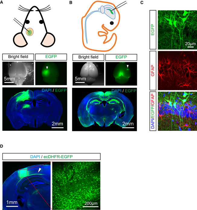
Figure EV1. Comparative assessment of transgene expression by AAV injection into rodent brain with two different protocols.

- Schematic diagram of AAV injection into one side of somatosensory cortex with cranial window in adult mouse brain. EGFP expression was introduced under control of synapsin promoter. Spatial distribution of EGFP was analyzed in whole brain (upper) and coronal brain slices (lower). Note that EGFP expression was highly restricted to injection site in this protocol. Arrowhead indicates AAV injection site.
- Schematic diagram of AAV injection into one side of lateral cerebral ventricle at postnatal day 0. Spatial distribution of EGFP was analyzed in whole brain (upper) and coronal slices (lower). Note that high‐level expression of EGFP is observed in circumferentially arranged area of the ventricle system including corpus callosum, hippocampus, and parietal cortex.
- Neuron‐specific expression of EGFP under control of synapsin promoter is verified by immunostaining using anti‐GFAP antibody. Representative images of neurons (green) and astrocytes (red) with DAPI staining (blue) in hippocampal CA1 captured by confocal microscopy are displayed.
- AAVs encoding ecDHFR‐EGFP were unilaterally injected into somatosensory cortex. Enriched neuronal expression of ecDHFR‐EGFP at injection site was assessed by fluorescence imaging of brain slices. Note arrowhead indicates that ecDHFR‐EGFP was also weakly distributed to axonal fibers derived from neurons of injection site.
