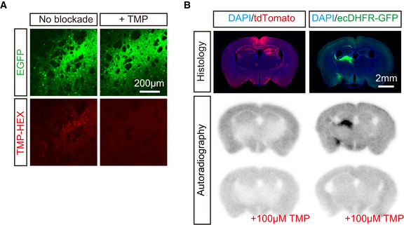
Figure EV3. In vitro validation of TMP‐HEX and [18F]FE‐TMP for ecDHFR.

- Representative images of striatal neurons expressing high‐level ecDHFR‐EGFP in fixed brain slices labeled with TMP‐HEX. Note that incubation with excess amount of non‐labeled TMP markedly inhibits fluorescence labeling.
- In vitro autoradiography of mouse brain sections with [18F]FE‐TMP. Expression of transgenes in samples collected from mice treated with control and ecDHFR‐EGFP vectors is fluorescently visualized with DAPI counterstaining (upper). Representative images of in vitro autoradiography using [18F]FE‐TMP demonstrate that this radioligand specifically labels brain areas overexpressing ecDHFR‐EGFP but not tdTomato (middle), and that this radioligand binding to putative ecDHFR is profoundly blocked by an excess amount of non‐labeled TMP (lower).
