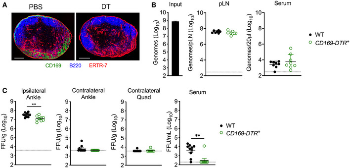
Figure 5. Macrophages in the dLN are not required for CHIKV accumulation in the dLN or limiting viral dissemination.

- CD169‐DTR+ mice were treated i.p. with PBS or DT prior to collection of the popliteal LN. Frozen LN sections were stained for CD169+ macrophages (green), B220+ B cells (blue), or ERTR‐7+ stromal cells (red). Image shown is representative of three biological replicates. Scale bars: 200 μm.
- WT or CD169‐DTR+ C57BL/6 mice were treated i.p. with DT prior to s.c. inoculation in the left‐rear footpad with 108 particles of WT CHIKV. Viral genomes in the dLN and serum at 2 hpi were quantified by RT–qPCR. Mean ± SD. Data are pooled from two experiments, n = 8. Mann–Whitney test; P > 0.05.
- WT or CD169‐DTR+ C57BL/6 mice were treated i.p. with DT prior to s.c. inoculation in the left‐rear footpad with 103 PFU of CHIKV. Infectious virus at 24 hpi was quantified by FFA. Mean ± SEM. Data are pooled from two experiments, n = 9–10. Mann–Whitney test; **P < 0.01.
